Dr. Takushi Namba, Associate Professor at Kochi University in Japan, discusses a research paper he co-authored that was published by Aging (Aging-US) in Volume 14, Issue 19, entitled, “Lotus germ extract rejuvenates aging fibroblasts via restoration of disrupted proteostasis by the induction of autophagy.”
Behind the Study is a series of transcribed videos from researchers elaborating on their recent studies published by Aging (Aging-US). Visit the Aging (Aging-US) YouTube channel for more insights from outstanding authors.
—
Hi. I am Takushi Namba, Associate Professor at Kochi University in Japan. Today, I will be sharing with you a recent published in Aging entitled, “Lotus germ extract rejuvenates aging fibroblasts via restoration of disrupted proteostasis by the induction of autophagy.”
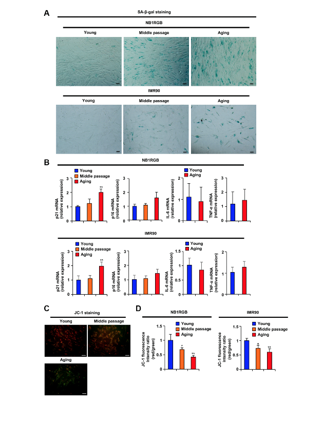
Cellular senescence induces multiple dysfunction of the cellular organism and slowly disrupts cellular homeostasis. First, we made different stages of proliferating and replicative senescence for human diploid fibroblasts and examined the expression level of aging-related markers. In these results, or this result, mitochondrial disfunction induced earlier the induction of DNA damaged signaling and SASP formation in the senescent fibroblast in early stage of cellular senescence. In this study, we used the senescent fibroblast.
It’s been previously reported that mitochondrial function is decreasing in the aging process, and mitochondrial dysfunction disrupts cellular homeostasis. Thus, we hypothesized that the activating mitochondria inhibits disrupt of cellular homeostasis in early stages of senescent fibroblasts.
We tried to screen for 75 plant extracts which are already used for humans as cosmetic or food materials, that increased mitochondrial transmembrane potential. Among these, lotus germ extract number 51 increased mitochondrial transmembrane activate potential the most in senescent fibroblasts. Next, we examined whether lotus germ extract affects SA-beta-gal activity in senescent fibroblasts. The number of SA-beta-gal-positive cells was decreased by the treatment of lotus germ extract, dose-dependent manner. The lesson is that lotus germ extract might suppress cell senescence phenotype.
To further understand the effect of lotus germ extract within cellular functions, we performed transmission electron microscope to understand any change in the cellular or around near structure. Lotus germ extract treatment-induced autolysosome-like structures show as red arrows in senescent fibroblasts.
We first examined whether lotus germ extract treatment affects autophagy induction and induced autophagy rates of reactivation of mitochondria and the suppression of aging phenotype. Senescent fibroblasts were treated with or without siRNA for ATG7 and/or lotus germ extract, and autophagy marker LC3-II levels were monitored. Treatment of lotus germ extract increased LC3-II protein expression and ATG7 knockdown by siRNA silencing. Suppressing lotus germ extract induced LC3-II protein expression. ATG7 knockdown significantly suppressed lotus germ extract-induced upregulation of mitochondrial transmembrane activate potential and the number of SA-beta-gal positive cells in senescent cells. These results indicated that lotus germ extract-induced autophagy has an important role in the activation of mitochondria.
Next, we focused on collagen production of aging fibroblasts in 2-D culture or 3-D culture. The production of total collagen protein was significantly increased by lotus germ extract treatment into the culture. We [inaudible] our three-dimensional cultural assay using autocollagen gel to evaluate its function of aging fibroblasts in the environment that more closely simulated in-people cultures. In tjhe process, staining assay reveals that collagen production increased in three-dimensional culture after treatment with lotus germ extract, indicating that lotus germ extract treatment leads toward extracellular matrix-remodeling activity in aging fibroblasts.
We performed several assays and then identified DAPK1 as a central role in lotus germ extract-induced autophagy. DAPK1 expression was suppressed by aging process-dependent epigenetic changes, which reduced histone acetylation and methylation. The treatment of lotus germ extract in senescent fibroblasts recovered epigenetic changes with induced histone acetylation and methylation, then a prevention of expression of DAPK1. Beclin1 and Bcl-2 complex suppressed Beclin1 activity. Upregulated DAPK1 also relates Beclin1, which is an active form, and then induced autophagy.
In this study, we identified that lotus germ extract activates mitochondria function by induced autophagy via DAPK1-Beclin1 similarly. Altogether, lotus germ extract is promising as a new anti-aging material and the induction of autophagy via DAPK1 is important as a new target in reactivation of mitochondria function in early stage of senescent cells.
An important point of this study, in the early stage of aging, the expression of certain proteins were decreased and aggregates of waste products were observed in the cells. The mitochondrial function was inferred by this disrupted proteostasis. As proposed in this study, the induction of autophagy, which reduced these waste products, is important to inhibit the progress of aging, and may also be involved in inhibition of aging.
Now we intend to explore how proteostasis is disrupted and how to maintain proteostasis during the aging process. Thank you for watching.
Click here to read the full study published by Aging (Aging-US).
AGING (AGING-US) VIDEOS: YouTube | LabTube | Aging-US.com
—
Aging (Aging-US) is an open-access journal that publishes research papers bi-monthly in all fields of aging research and other topics. These papers are available to read at no cost to readers on Aging-us.com. Open-access journals offer information that has the potential to benefit our societies from the inside out and may be shared with friends, neighbors, colleagues, and other researchers, far and wide.
For media inquiries, please contact [email protected].